

Cranial Phantoms
Cranial and Neurosurgical Training Models with Realistic Tissue Response
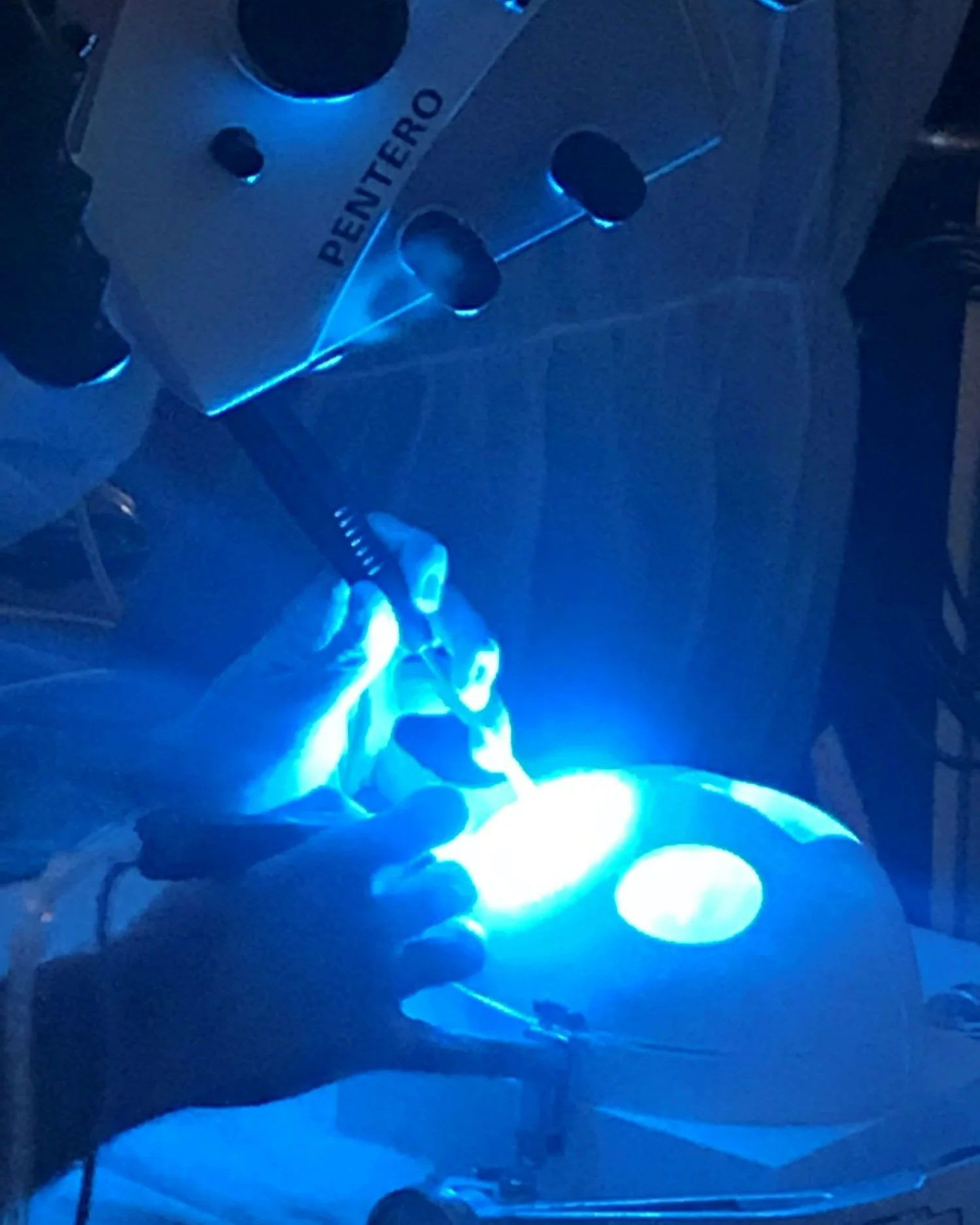
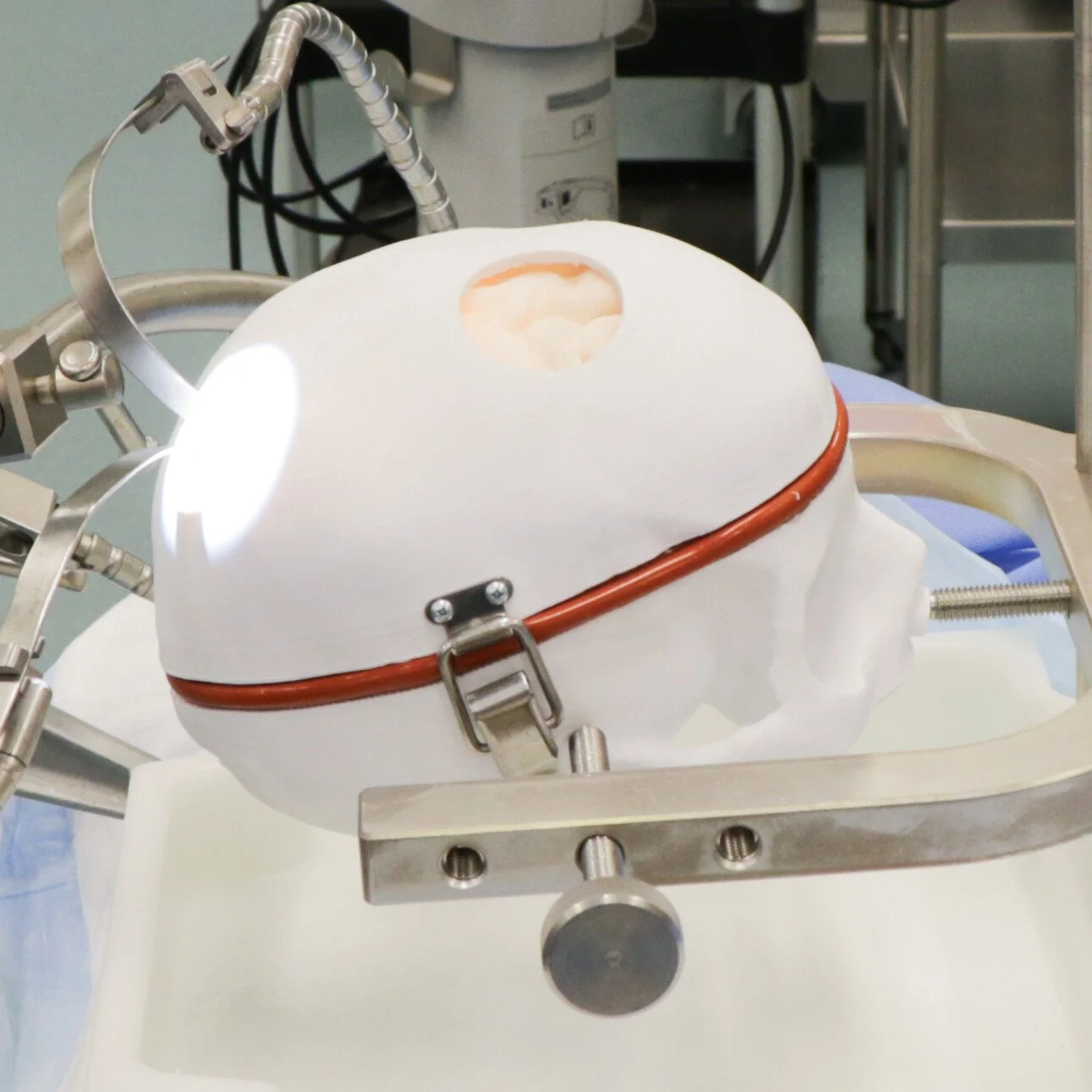
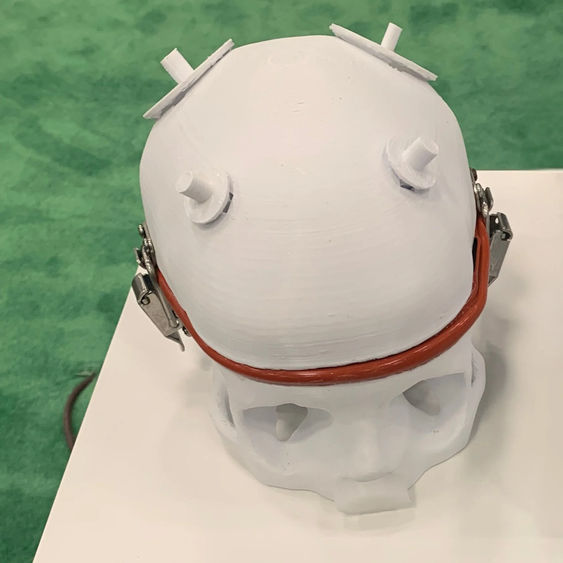
Viomerse cranial phantoms are high-fidelity anatomical models developed for neurosurgical training, procedural simulation, and medical device evaluation involving the skull and brain. These realistic synthetic cadavers incorporate anatomical elements that respond authentically to drilling, cutting, suturing, electro-cautery and manipulation while maintaining accurate imaging characteristics. Used by surgeons, educators, and medical device companies, Viomerse cranial phantoms support hands-on training and validation for tumor visualization, tumor resection, craniotomy, neuro-navigation, and skull-base procedures.

Multiple Options - Resectable, Nonresectable, and Replacement Brain
Resectable: Soft hydrogel brain with skull casing.
Synthetic hydrogel brain with 3 resectable tumors and a tumor cavity within the cerebral cortex, contained within a plastic skull base with ports of entry for tumor access. Retraction devices not required for use. Tumors are compatible with fluorescent imaging; phantom includes MRI DICOM.
Nonresectable: Durable rubber brain with skull casing.
Phantom intended for demonstration of fluorescent microscopy. Includes tumors simulating imaging with 5-ALA, fluorescein, and a synthetic blood vessel with ICG. Phantom is reusable, not intended for procedural simulation, and made from durable rubber.
Replacement Brain: Soft hydrogel brain without skull casing.
Synthetic hydrogel brain with 3 resectable tumors and a tumor cavity within the cerebral cortex. Does not contain the plastic skull base or MRI DICOM files; serves as a replacement brain for a previously purchased Cortical Brain Tumor Phantom.
2 Options: Full Procedure and Partial Procedure
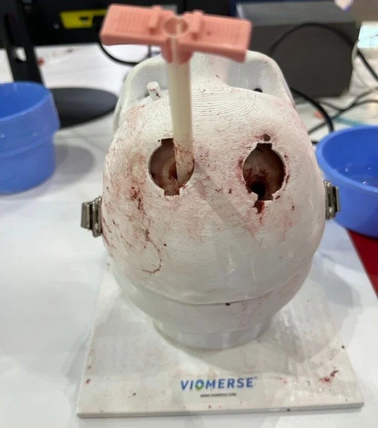
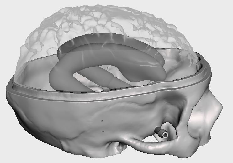
Full Procedure: Synthetic brain with 4 resectable tumors within the thalamic and parietal occipital regions of the brain, contained within a plastic skull base with ports of entry for tumor access. Requires retraction for tumor access. Synthetic skull requires burr hole procedure for access. Tumors are compatible with fluorescent imaging; phantom includes MRI DICOM files.
Partial Procedure: Synthetic brain with 4 resectable tumors within the thalamic and parietal occipital regions of the brain, contained within a plastic skull base with ports of entry for tumor access. Requires retraction for tumor access. Tumors are compatible with fluorescent imaging; phantom includes MRI DICOM files.
3 Options: Full Procedure, Partial Procedure, Refillable
Full Procedure: Includes plastic cranial base in the supine position, scalp, a frontal bone with synthetic cortical and cancellous layers, dura mater, and a realistic hydrogel brain containing 2 hematomas. Includes CT scan DICOM file.
Partial Procedure: Includes synthetic cranial base in the supine position, plastic frontal bone, and a realistic hydrogel brain containing 2 hematomas. Includes CT scan DICOM file.
Refillable: Includes plastic cranial base in the supine position with a plastic scalp that has 2 preformed access pathways to two chambers. The brain included is durable and intended for re-use. Synthetic blood clot refill kit available for $195.
2 Options: Full Procedure and Partial Procedure
Full Procedure: Includes a synthetic hydrogel scalp and synthetic skull requiring identification of Kocher’s point and burr hole drilling. Phantom is attached to a fluid source. Each model has a hydrogel brain and can tolerate 3 to 4 punctures per ventricle with fluid efflux.
Partial Procedure: Includes a synthetic silicone scalp and plastic skull with pre-made burr holes at Kocher’s point, attached to a fluid source. Each model has a hydrogel brain and can tolerate 3 to 4 punctures per ventricle with fluid efflux.
Cranial phantom intended for repeated ultrasound imaging of the brain. Hydrogel brain includes tumor, resected tumor cavity, ventricles, falx cerebri, and pituitary lesion visible through transsphenoidal passage.