

Dr. Nelson Stone, Urologist and Radiation Oncologist
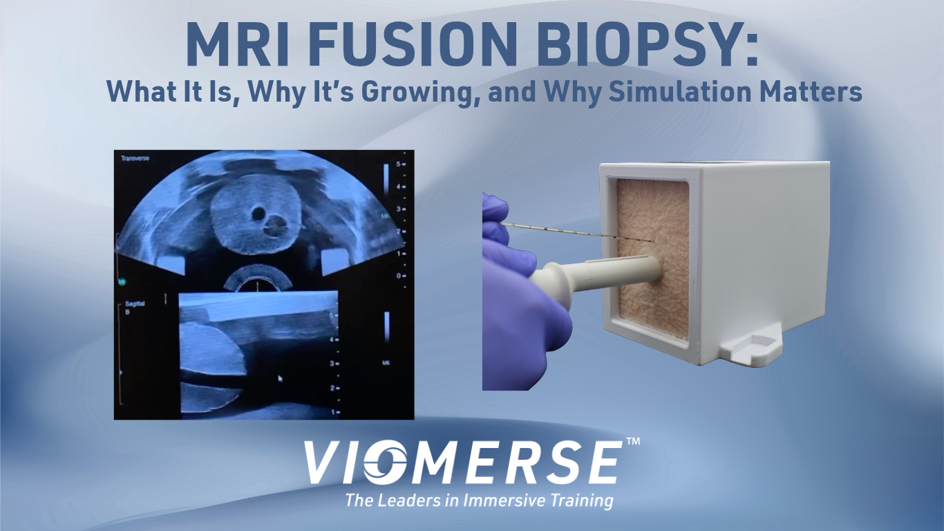
MRI fusion biopsy is becoming an increasingly important technique in prostate cancer diagnosis. By combining MRI information with real-time ultrasound, urologists can more precisely target suspicious areas of the prostate during biopsy.
As more urology practices adopt dedicated MRI-ultrasound fusion systems, there is a growing need for effective fusion biopsy training. Physicians need to understand not only how to perform a prostate biopsy, but also how to interpret MRI findings, register imaging datasets, navigate to a target, and operate specialized fusion equipment.
Realistic simulation can help physicians develop these skills before performing the procedure on patients.
Viomerse has developed fusion prostate biopsy phantoms specifically to support this type of hands-on MRI fusion biopsy education.
What Is an MRI Fusion Biopsy?
An MRI fusion biopsy, also called a prostate fusion biopsy or targeted prostate biopsy, combines information from magnetic resonance imaging (MRI) with live ultrasound imaging.
For men with an elevated PSA (prostate-specific antigen), a high-risk profile, or an abnormal digital rectal examination, an MRI may be used to identify suspicious areas within the prostate. These findings are commonly reported using the PI-RADS (Prostate Imaging Reporting and Data System) classification.
When a suspicious lesion is identified, the urologist may perform a targeted biopsy to obtain tissue samples from that specific area.
The challenge is that a lesion identified on MRI may not be clearly visible on live ultrasound. MRI fusion technology addresses this challenge by helping the physician correlate the MRI findings with the real-time ultrasound image.
How MRI Fusion Biopsy Works
A typical MRI fusion biopsy workflow involves several steps.
First, the physician reviews the patient's prostate MRI and identifies the suspicious lesion or target.
The MRI data can then be loaded into a dedicated fusion biopsy system. During the procedure, the physician acquires live ultrasound images of the prostate.
The fusion system registers the MRI data with the ultrasound images, allowing the physician to visualize the MRI-defined target in relation to the live ultrasound anatomy.
The physician can then use the fused images to guide the biopsy needle toward the target.
This workflow requires skills that go beyond those used for a conventional ultrasound-guided prostate biopsy.
Cognitive Fusion vs. Targeted MRI Fusion Biopsy
There are different approaches to combining MRI information with ultrasound during prostate biopsy.
Cognitive Fusion
With cognitive fusion, the physician reviews the MRI and mentally translates the location of the suspicious lesion to the live ultrasound image.
In other words, the urologist looks at the MRI, looks at the ultrasound, and uses their knowledge of prostate anatomy to estimate where the MRI-identified lesion should be located.
Cognitive fusion does not require dedicated MRI-ultrasound fusion equipment, but it relies heavily on the physician's ability to interpret the MRI and correlate it with the ultrasound anatomy.
Software-Assisted MRI Fusion
A more technology-driven approach uses dedicated fusion biopsy equipment to register the MRI with live ultrasound.
This allows the physician to work with a combined image and more directly target the area identified on MRI.
For physicians accustomed to conventional ultrasound-guided prostate biopsy, learning this workflow introduces an additional technical skill set—including image registration, target identification, system navigation, and needle guidance.
That makes hands-on practice an important part of MRI fusion biopsy training.
Why Is MRI Fusion Biopsy Becoming More Popular?
Several developments are contributing to increased interest in targeted prostate biopsy.
MRI has become an increasingly important tool for identifying suspicious prostate lesions. Rather than relying solely on systematic biopsy sampling, physicians can use MRI findings to help identify areas that warrant targeted tissue sampling.
At the same time, MRI-ultrasound fusion technology has continued to develop, with multiple medical technology companies offering dedicated systems for targeted prostate biopsy.
Changes in reimbursement in the United States beginning in January 2026 have also increased interest among urology practices evaluating dedicated fusion biopsy technology.
As more practices adopt these systems, there is a corresponding need for physicians to develop proficiency with the equipment and workflow.
Why MRI Fusion Biopsy Requires Specialized Training and Tools
Performing a traditional ultrasound-guided prostate biopsy and performing a software-assisted MRI fusion biopsy involve overlapping skills, but they are not identical procedures.
A physician learning an MRI fusion system may need to become comfortable with:
Reviewing and interpreting prostate MRI findings
Identifying and defining biopsy targets
Acquiring and manipulating ultrasound images
Registering MRI and ultrasound datasets
Navigating the fusion software
Locating the target within the fused image
Guiding the biopsy needle to the intended target
Understanding the workflow of a particular fusion platform
The ability to practice these steps repeatedly can help physicians become familiar with the technology before incorporating it into their clinical practice.
How an MRI Fusion Biopsy Phantom Supports Hands-On Training
Medical simulation provides an opportunity to practice procedures in a controlled, repeatable environment.
A prostate biopsy phantom can allow physicians to perform simulated biopsies without the pressure of working directly with a patient.
For MRI fusion training, however, a conventional ultrasound phantom may not be enough. The training model needs to support the imaging workflow associated with targeted biopsy.
An MRI fusion biopsy phantom can provide a simulated target within a prostate model while also supplying an MRI dataset that can be incorporated into the fusion system.
This allows learners to practice the same basic workflow they will encounter during an MRI fusion biopsy.
Viomerse MRI Fusion Biopsy Phantoms for Prostate Biopsy Training
Viomerse has developed specialized fusion prostate phantoms to help urologists, educators, and medical device teams train for MRI fusion biopsy procedures.
Each phantom is designed to support a workflow that combines MRI information with live ultrasound.
The phantom is supplied with an MRI DICOM file, allowing learners to practice importing and working with MRI data as part of the fusion biopsy process.
Because the model is encased and easily transportable, it can be used in a variety of educational and training environments, including:
Urology training programs
Simulation centers
Medical education workshops
Physician skills laboratories
Device demonstrations
Fusion biopsy system training
Hands-on continuing medical education
The phantom can also be used with many modern fusion biopsy platforms, providing flexibility for institutions and medical device companies conducting hands-on training.
Practice MRI Fusion Biopsy Before Treating Patients
One of the primary advantages of simulation is the ability to practice repeatedly.
A physician learning a new fusion biopsy system can work through the imaging workflow, practice registration, identify simulated targets, and perform needle guidance without using a patient as the training environment.
Instructors can also use the phantom to demonstrate the procedure and allow learners to repeat specific steps until they become more comfortable with the equipment.
For institutions introducing a new MRI fusion biopsy platform, this creates an opportunity to provide hands-on training before clinical implementation.
Preparing Urologists for the Future of Targeted Prostate Biopsy
As prostate MRI becomes increasingly integrated into prostate cancer diagnosis, targeted biopsy techniques are becoming an important part of the urologist's toolkit.
The growth of dedicated MRI-ultrasound fusion systems creates new opportunities for more targeted prostate biopsy—but it also creates a need for effective physician education.
MRI fusion biopsy training should give physicians an opportunity to learn the technology, practice the workflow, and build familiarity with the equipment before performing targeted biopsies on patients.
Viomerse fusion prostate phantoms are designed to provide that hands-on training environment.
By combining a realistic prostate model with an associated MRI dataset, the Viomerse phantom allows physicians and educators to simulate the MRI fusion biopsy workflow in a controlled and repeatable setting.
Frequently Asked Questions About MRI Fusion Biopsy
What is an MRI fusion biopsy?
An MRI fusion biopsy combines prostate MRI data with real-time ultrasound imaging to help a urologist target suspicious areas of the prostate during biopsy. The MRI identifies areas of concern, while the fusion system helps the physician correlate those findings with the live ultrasound image and guide the biopsy needle toward the target.
What is the difference between cognitive fusion and MRI fusion biopsy?
With cognitive fusion, the urologist reviews the MRI and mentally correlates the location of the suspicious lesion with the live ultrasound image. An MRI fusion biopsy using dedicated software and equipment digitally registers the MRI with the ultrasound image, providing a more formal image-fusion workflow for targeting the lesion.
Why is MRI fusion biopsy becoming more popular?
The increasing use of prostate MRI, continued development of dedicated MRI-ultrasound fusion systems, and changes in U.S. reimbursement beginning in 2026 have contributed to growing interest in targeted prostate biopsy. As more urology practices adopt fusion biopsy technology, physicians also need opportunities to develop proficiency with the equipment and workflow.
How do physicians learn MRI fusion biopsy?
Learning MRI fusion biopsy requires more than understanding the underlying technology. Physicians need hands-on experience with the fusion system, including MRI registration, target identification, ultrasound imaging, system navigation, and needle guidance. Simulation with a realistic MRI fusion biopsy phantom allows physicians to practice these steps in a controlled and repeatable environment before performing the procedure on patients.
What is an MRI fusion biopsy phantom?
An MRI fusion biopsy phantom is a realistic training model designed to simulate the prostate biopsy workflow using MRI and ultrasound. Viomerse prostate phantoms are supplied with an MRI file and can be used with many modern fusion biopsy systems, allowing physicians, educators, and medical device teams to practice targeted biopsy techniques in a hands-on training environment.
Dr. Nelson Stone, Urologist and Radiation Oncologist